Showing posts with label hippocampus. Show all posts

Monday, August 26

Your Brain's Memory Resets


A new study from Cornell University reveals that sleep not only consolidates memories but also resets the brain’s memory storage mechanism. This process, governed by specific regions in the hippocampus, allows neurons to prepare for new learning without being overwhelmed. This insight opens potential pathways for enhancing memory and treating neurological disorders like Alzheimer’s and PTSD.


While everyone knows that a good night’s sleep restores energy, a new Cornell University study finds it resets another vital function: memory.

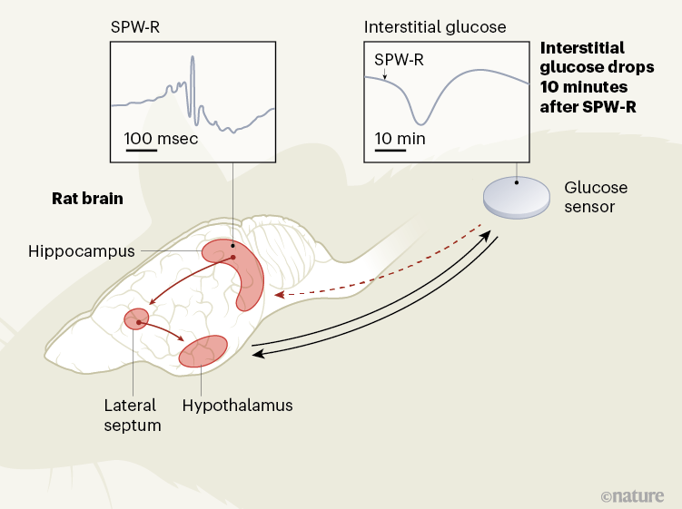
Learning or experiencing new things activates neurons in the hippocampus, a region of the brain vital for memory. Later, while we sleep, those same neurons repeat the same pattern of activity, which is how the brain consolidates those memories that are then stored in a large area called the cortex. But how is it that we can keep learning new things for a lifetime without using up all of our neurons?
Mechanisms of Memory Resetting

A new study published in the journal Science, finds at certain times during deep sleep, certain parts of the hippocampus go silent, allowing those neurons to reset.

“This mechanism could allow the brain to reuse the same resources, the same neurons, for new learning the next day,” said Azahara Oliva, assistant professor of neurobiology and behavior and the paper’s corresponding author.         READ MORE...


Thursday, August 19

Thought and Metabolism

To regulate adaptive behaviour, the brain relies on a continuous flow of cognitive and memory-related processes that require a constant energy supply. Weighing around 1,200 grams in women and 1,300 grams in men, on average, the brain consumes around 90 grams, or 340 kilocalories’ worth, of glucose per day, accounting for around half of the body’s glucose demand1,2

The tight integration of metabolic and cognition-related signals might aid the matching of the brain’s energy supply to its energy needs, by optimizing foraging behaviour and efforts to limit energy expenditure. 

The synchronization of glucose supply with brain activity has so far been considered a function of a structure called the hypothalamus, at the base of the brain. Writing in Nature, Tingley et al.3 provide evidence in rats for the role of another brain region, called the hippocampus, which is typically implicated in memory and navigation, in this equation (Fig. 1).


Figure 1 | Brain signals that regulate glucose levels in the body periphery. The hypothalamus in the brain helps to regulate glucose concentrations in the blood and in the interstitial fluid that surrounds cells in the body. This hypothalamic (feedback-mediated) regulation is activated, for example, during stress. Tingley et al.3 provide evidence in rats that another brain structure, the hippocampus, also regulates peripheral glucose concentrations. In the hippocampus, oscillatory patterns — called sharp wave-ripples (SPW-Rs) — emerge in the collective electrical potential across the membranes of neurons. They seem to signal, by way of a region called the lateral septum, to the hypothalamus to produce dips in interstitial glucose concentration about 10 minutes later. The feedback mechanism in this regulatory loop is unknown (dashed arrow). Given that hippocampal SPW-Rs are a hallmark of the reprocessing of previous experiences, they might thus control the brain’s energy supply during a ‘thought-like’ mode.

The hippocampus receives many types of sensory and metabolic information, and projections from neuronal cells in the hippocampus extend to various parts of the brain, including the hypothalamus. Thus, the hippocampus might indeed represent a hub in which metabolic signals are integrated with cognitive processes3

To examine this possibility, Tingley and colleagues recorded oscillatory patterns called sharp wave-ripples (SPW-Rs), reflecting changes in electrical potential across the cell membranes of neuronal-cell ensembles in the hippocampi of rats. They did this while using a sensor inserted under the skin of the animals’ backs to continuously measure glucose levels in the interstitial fluid surrounding the cells there.  READ MORE

Friday, July 23

Adult Brains


(Image caption: A 3-D animated image showing our synapse phagocytosis reporter in mouse hippocampus. Presynapses in green, astrocytes in white, and microglia in blue. Phagocytosed presynapses by glia were shown in red.)


Astrocytes Eat Connections to Maintain Plasticity in Adult Brains
Developing brains constantly sprout new neuronal connections called synapses as they learn and remember. Important connections — the ones that are repeatedly introduced, such as how to avoid danger — are nurtured and reinforced, while connections deemed unnecessary are pruned away. Adult brains undergo similar pruning, but it was unclear how or why synapses in the adult brain get eliminated.

Now, a team of KAIST researchers has found the mechanism underlying plasticity and, potentially, neurological disorders in adult brains. They published their findings in Nature.

“Our findings have profound implications for our understanding of how neural circuits change during learning and memory, as well as in diseases,” said paper author Won-Suk Chung, an assistant professor in the Department of Biological Sciences at KAIST. “Changes in synapse number have strong association with the prevalence of various neurological disorders, such as autism spectrum disorder, schizophrenia, frontotemporal dementia, and several forms of seizures.”

Gray matter in the brain contains microglia and astrocytes, two complementary cells that, among other things, support neurons and synapses. Microglial are a frontline immunity defense, responsible for eating pathogens and dead cells, and astrocytes are star-shaped cells that help structure the brain and maintain homeostasis by helping to control signaling between neurons. According to Professor Chung, it is generally thought that microglial eat synapses as part of its clean-up effort in a process known as phagocytosis.

“Using novel tools, we show that, for the first time, it is astrocytes and not microglia that constantly eliminate excessive and unnecessary adult excitatory synaptic connections in response to neuronal activity,” Professor Chung said. “Our paper challenges the general consensus in this field that microglia are the primary synapse phagocytes that control synapse numbers in the brain.”

Professor Chung and his team developed a molecular sensor to detect synapse elimination by glial cells and quantified how often and by which type of cell synapses were eliminated. They also deployed it in a mouse model without MEGF10, the gene that allows astrocytes to eliminate synapses. Adult animals with this defective astrocytic phagocytosis had unusually increased excitatory synapse numbers in the hippocampus. Through a collaboration with Dr. Hyungju Park at KBRI, they showed that these increased excitatory synapses are functionally impaired, which cause defective learning and memory formation in MEGF10 deleted animals.

“Through this process, we show that, at least in the adult hippocampal CA1 region, astrocytes are the major player in eliminating synapses, and this astrocytic function is essential for controlling synapse number and plasticity,” Chung said.

Professor Chung noted that researchers are only beginning to understand how synapse elimination affects maturation and homeostasis in the brain. In his group’s preliminary data in other brain regions, it appears that each region has different rates of synaptic elimination by astrocytes. They suspect a variety of internal and external factors are influencing how astrocytes modulate each regional circuit, and plan to elucidate these variables.

“Our long-term goal is understanding how astrocyte-mediated synapse turnover affects the initiation and progression of various neurological disorders,” Professor Chung said. “It is intriguing to postulate that modulating astrocytic phagocytosis to restore synaptic connectivity may be a novel strategy in treating various brain disorders.”